Clinical case
Clinical case: operation for L4L5 transforaminal and L5S1 interlaminar disc herniations
After a previous operation, the patient presents with sciatica to our spine specialist center, which causes continuous and disabling pain.
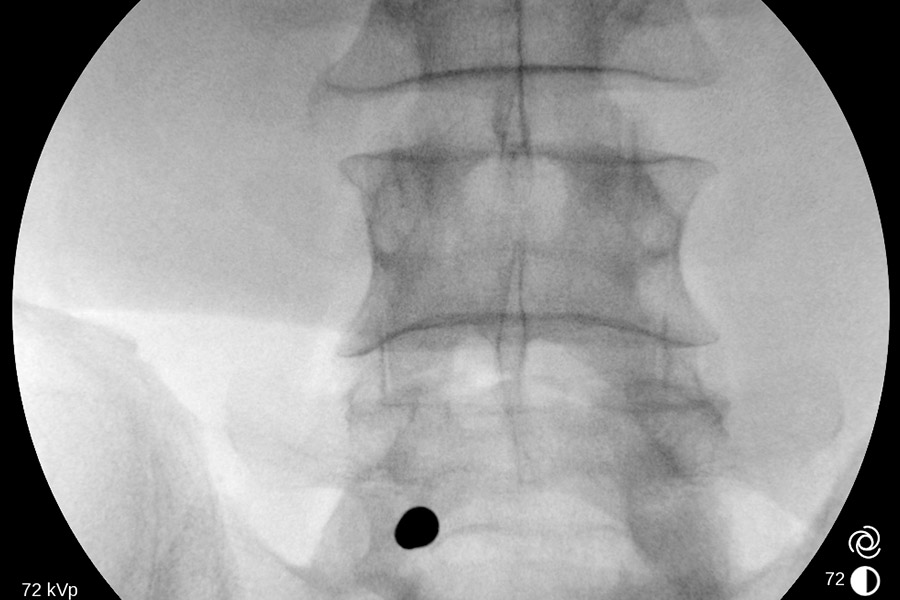
Preoperative
The patient had previously undergone surgery for an L4L5 disc herniation. After undergoing this surgery, his pathology improved significantly, but after a few months he developed sciatica similar to what he had before said operation. It was less intense but with continued pain.
Once the patient was studied thanks to the MRI images and clinical examination, it was observed that he had fibrosis in the area that had previously been operated on. This Fibrosis may be the cause of compression of the neurological system, causing sciatica. In addition, it was observed that there is also a herniated disc between the fifth lumbar vertebra and the sacrum. In this case they will address both levels to release these neurological structures.
Operation
You opt for the transforaminal approach accessing the canal. In the video you can see the herniated disc that appears because it is not in its natural location, so the fibers of the disc are not structured.
Little by little the tissue coagulates and then small incisions are made to remove the hernia more easily. One of the medical devices used is a coagulator: a high-precision radiofrequency terminal that allows tissues to be retracted and made smaller and smaller. It has several functions: the aforementioned coagulation serves to close the vessels that are bleeding, while vaporization is used to eliminate tissues. Then we reach the disc material, which will be progressively removed.
In this case, the material is quite large, so it is more difficult to remove it in one piece, so it will be extracted little by little, in turn decompressing the neurological structures. This material is also found within the canal itself, compressing its neurological structures. Furthermore, it is anchored to fibrosis since this patient had already been operated on with another approach technique.
As we pull the disc material, as the adhesions and fibrosis break down, the blood vessels begin to bleed a little, so they progressively coagulate to prevent the formation of fibrosis in the future and to have optimal vision throughout the operation . The material is anchored to the fibrous tissue due to the patient's previous open surgery.
Postoperative
This procedure does not last long. It continues to be cleaned and coagulated little by little until the structures are completely freed, creating a fibrous layer that allows the formation of a type of membrane that protects it from future hernias.
In addition, this surgical technique allows us to check if there are any loose fragments that would also need to be released.